Friday, 6 March 2020

Why is the human sex ratio 1:1 at birth?

Having found a dramatic shift in sex ratio at birth in one species of mammal, I have for decades kept an eye on discussions as to whether the human sex ratio at birth differs in some circumstances from 1 male: 1 female. Mammals in theory have two ways of controlling the sex ratio at birth: (i) at conception, or (ii) later by selectively reducing the number of young in utero. We found the latter mechanism at work in the guinea-pig. Since human litter size usually equals the  number of eggs and rarely exceeds 1 and since gestation is relatively long, it always appeared that if there were to be any maternal or paternal genetic control of the sex of the offspring it would have to be at the time of conception, rather than by selective death and reabsorption of embryo or fetus.

In the 1980s I once did the experiment of asking biologists from different disciplines why they thought the human sex ratio is 1:1. The reproductive biologists replied that it was just the result of random segregation of the sex chromosomes. The sex of a human offspring depends on whether it inherits and X or a Y chromosome from its father. Random segregation will, on average, result in a 1:1 ratio. By contrast, the evolutionary biologists said that the sex ratio is explained by Sir Ronald Fisher’s Principle: with the sex of a subject to genetic variation, the the sex ratio will always stabilise at 1:1. I will not repeat the simple explanation that can be found here.


Shifts of the of sex ratio at birth in various animals has been explained in terms of the Trivers-Willard hypothesis: parents which possess a heritable trait that benefits the lifetime reproductive success of one sex will bias the sex ratio towards that sex. There have been various claims in studies of human populations along these lines; for example, male-biased sex ratios in taller, wealthier, high status parents with the offspring likely to be more successful in competition for a mate. However, such claims have been controversial because of the statistical methods used and the results have often not borne out when larger samples were taken from the population.

Both the Fisher Principle and Trivers-Willard rely on there being genetic variation in the sex ratio, in other words that a bias towards one sex or the other is heritable. Thus a key test is to look for heritability in a very large human population. On standard scale of 0 to 1, ‘0’ denotes that a trait is not heritable while ‘1’ all differences in a trait can be explained entirely by genetic variation. Many traits fall somewhere between those two extremes.

But what does determine human sex ratio? It would be predicted if sex ratio is a heritable trait then Fisher’s Principle would apply. By contrast, if it is not heritable then simple Mendelian segregation of the sex chromosomes would suffice as an explanation.

Over the years there have been all sorts of suggestions and claims that the tendency in a family to produce offspring completely or partially biased to one sex is hereditary, and that particular genes could be involved. However, these conclusions have been criticised because the sample sizes were small and the statistical inferences drawn were invalid.

A recent, important paper has tackled the problem by using data from the entire population born in Sweden in and after 1932. That was 3,543,243 individuals and their 4,753,269 children. The results of the analysis were clear. There was no evidence of heritability at all. The calculated heritability was 0. In other words, there was no need to invoke Fisher’s Principle since with no heritability there can be no Fisher.

The authors summed up their results:

In sum, all of our results are consistent with the simple explanation that variation in offspring sex ratio in humans is due to unbiased Mendelian segregation of sex chromosomes during spermatogenesis and unbiased fertilization. The slight excess of male births is likely to be due to a general difference in survival of male and female embryos in the womb, the reasons for which are not yet understood.


Looks like my reproductive biology colleagues were right. Pity I can’t tell most of them; they are long dead.

The question now, of course, is whether the same conclusion, that the human sex ratio at conception is simply the outcome of random segregation of the sex chromosomes, applies to other or to all mammals? And are the statistically-robust demonstrated shifts in sex ratios at birth in some species and in certain environmental conditions all the result of differential loss of embryos and fetuses in utero? I shall permit myself to guess that the answers are ‘Yes’ and ‘Yes’ even though, as with previous human studies, there have been claims that the answer to the first question is ‘No’.


Zietsch BP, Walum H, Lichtenstein P, Verweij KJH, Kuja-Halkola R. 2020 No genetic contribution to variation in human offspring sex ratio: a total population study of 4.7 million births. Proceedings of the Royal Society B 287: 20192849. http://dx.doi.org/10.1098/rspb.2019.2849

Peaker M, Taylor E. 1996. Sex ratio and litter size in the guinea-pig. Journal of Reproduction and Fertility 108, 63-67.


Wednesday, 4 March 2020

Fluorescent chameleons, salamanders, newts and frogs...and a budgerigar

An increasing number of animals are being found to fluoresce.  Fluorescence happens when a chemical fluorophore absorbs of shorter-wavelength light and them emits some of the absorbed energy as light at a longer-wavelength. In some biological examples, blue light is absorbed and green light is emitted

Why animals fluoresce is the subject of active research. Does the change to a more noticeable colour mean that fluorescence is sometimes involved in signalling to other animals of the same sex, or of  different sex, or of the same species, or to a predator, or to prey? Is it assisting in camouflage, the fluorescent emission matching, say, the emission from plants in the background?

Perception of fluorescence depends on the properties of the photoreceptors of the animal exposed to the fluorescence, so that in the case of proposed signalling between animals of the same species, it would be a requirement to demonstrate that the visual pigments of the eyes of that species can actually pick up the colour.

There is, of course, the possibility that there is no function of the fluorescence at all, that it is just a by-product of a particular molecule used as a pigment or for some other purpose in the skin or other tissue. This is the same argument as to why some creatures from the depths of the sea have bright colours; the production of brightly coloured molecules used by the animal are simply not selected against since there is no predator present that can see them.

A very well argued paper appeared in 2017 in Philosophical Transactions of the Royal Society by Justin Marshall (University of Queensland) and Sonke Johnsen (Duke University, North Carolina). The authors explained the background to explaining any function of fluorescence in terms of communication between individuals. They set out criteria that must be satisfied in order for such a role for fluorescence to be accepted. The Budgerigar, Melopsittacus undulatus, they found from descriptive and experimental studies, fulfilled all the criteria. In short, both males and females prefer to associate with potential mates that fluoresce.


Fluorescence characteristics of the budgerigar
(a,b) White light and UV fluorescent excitation
photographs of front and back of head showing
fluorescent cheek and crown feathers
from Marshall & Johnsen 2017


More recent studies have shown fluorescence from the bony tubercles of the skull of many species of chameleon—just one more feature added to the list of properties of those extraordinary animals. The phenomenon is particular evident in those chameleons living in humid forests ‘known to have a higher relative component of UV light’. The blue light emitted would be in sharp contrast to the brown and green colours reflected by the surroundings.


from Prötzel et al 2018


Even more recently came photographs showing fluorescence in a number of amphibians, this time a green emission from blue light (with some species showing emission in response to UV (ultraviolet)).


from Lamb & Davis 2020


The accumulating evidence for fluorescence in a wide range of animals (and plants) raises so many questions as to the molecular mechanisms involved and possible functions, that the only conclusion at present is the inevitable: more research is required.

In the meantime enjoy the pictures.


Lamb JY, Davis MP. 2020. Salamanders and other amphibians are aglow with biofluorescence. Scientific Reports 10, 2821. https://doi.org/10.1038/s41598-020-59528-9 

Marshall J, Johnsen S. 2017 Fluorescence as a means of colour signal enhancement. Phil. Trans. R. Soc. B 372: 20160335. http://dx.doi.org/10.1098/rstb.2016.0335 

Prötzel D, Heß M, Scherz MD, Schwager M, van’t Padje A, Glaw F. 2018. Widespread bone-based fluorescence in chameleons. Scientific Reports 8, 698. DOI:10.1038/s41598-017-19070-7 

Tuesday, 3 March 2020

Peter Charles Howard Pritchard 1943-2020. An encyclopaedic knowledge of chelonians and their conservation. How did he start out?

The study and conservation of chelonians—turtles, terrapins and tortoises—has lost a major champion with the death on 25 February of Peter Pritchard at the age of 76. He and his Chelonian Research Institute were better known in the U.S.A. and the lands where chelonians breed than in his native United Kingdom. An appreciation of his work, influence and importance can be read on the website of Turtle Conservancy.


From Turtle Conservancy's website


I never met Peter Pritchard and often wondered how he had become interested in reptiles. Then I read an appreciation (on what is now a dead link) he had written of Angus d’Albini Bellairs (1918-1990):

Angus was the first herpetologist I ever met. He was the immediate successor to my father as Reader in Anatomy at St. Mary’s Hospital, my father (Dr. J. J. Pritchard*) having been appointed Professor of Anatomy at Queen’s University in Belfast, Northern Ireland the preceding year, and this connection gave me free access to a man I regarded as an intellectual hero.  My (signed) copy of his 1957 book, simply named Reptiles, is dated April 5 1958, when I was just 14 years old, and just a few years later I started to write a book of my own, which I ambitiously entitled Living Turtles of the World.  Despite the clearly schoolboyish flavor of this early draft, not to mention the lack of personal field experience and shortage of library access, Angus introduced me to the concept of peer review (although we were not exactly peers), and he read the whole thing, making gentle suggestions in pencil wherever he saw fit. 
Whenever I was in London, I would find my way to the dusty chambers of St. Mary’s (made famous by Sir Alexander Fleming), and knock on Angus’ door for a conversation on the subject of mutual interest, namely herpetology.  At such times, he would always open a bottle of sherry and bring some small-size laboratory beakers from which we would drink it, as he urged me to pursue an experimental approach to herpetology, by means such as studying underwater respiration in softshell turtles, or scute regeneration in chelonians.  (Somehow, I never became “experimental”, instead concentrating on natural history, taxonomy, skeletal anatomy, and conservation aspects).
Angus also introduced me to other herpetologists, including Miss Grandison, the Keeper of Zoology at the British Museum, a lady who had seemed rather remote and doctrinaire when I wrote to her, but was more like a favorite auntie once encountered in person.  I was also privileged, through her, to meet J. C. Battersby, on his very last day before retiring in 1961; he had been a “boy attendant” of G. A. Boulenger, no less, appointed in 1916.
Angus introduced me to the bizarre militaristic hierarchies at the London Zoo (Regents’ Park), where the “gentlemen officers” (the Curators etc) lorded it over the non-commissioned ranks (head keepers and below), only the former being admitted to such places as the Fellows’ Restaurant.  He himself was, of course, “top of the heap,” a scholar and a gentleman, although unpaid in status as “honorary herpetologist,” and I think his extensive wartime military experience was what prompted him to refer to the Reptile House staff as his “sergeant major,” his “corporal,” etc… 

And that’s how it all began, although his interest in animals was first piqued by being taken as a small boy to London Zoo. Even though his first degree was in chemistry at Oxford, he moved to Florida to work for his Ph.D. with Archie Carr (1909-1987), the doyen of research and conservation of turtles.

The book Pritchard referred to was his Living Turtles of the World, published in 1967—when he was 24—by the infamous Herbert R. Axelrod’s TFH Publications. A completely rewritten survey of the world’s chelonians stretching to nearly 900 pages, also published by TFH, appeared as Encyclopedia of Turtles in 1979. Axelrod himself took a number of the photographs. I have a copy—still a useful reference—on my shelves.

The following video on, and including Peter Pritchard, appeared in 2016.


*Peter Pritchard’s father. John Joseph (Jack) Pritchard. was born in 1916 in Adelaide, South Australia. By the age of 19 he had a first degree and arrived in Oxford on a Rhodes Scholarship to work at Oxford in the Department of Physiology. After becoming medically qualified in London he moved into anatomy, first at UCL, then St. Mary’s before moving to Belfast as Professor. His research was on bone growth and repair, a subject the reader will notice of considerable interest and importance in the life of chelonians. He died in 1979, aged 63.


Friday, 28 February 2020

Chameleons I have kept and chameleons I have seen in the wild

This Jackson's Chameleon was the second chameleon I
kept ca 1960
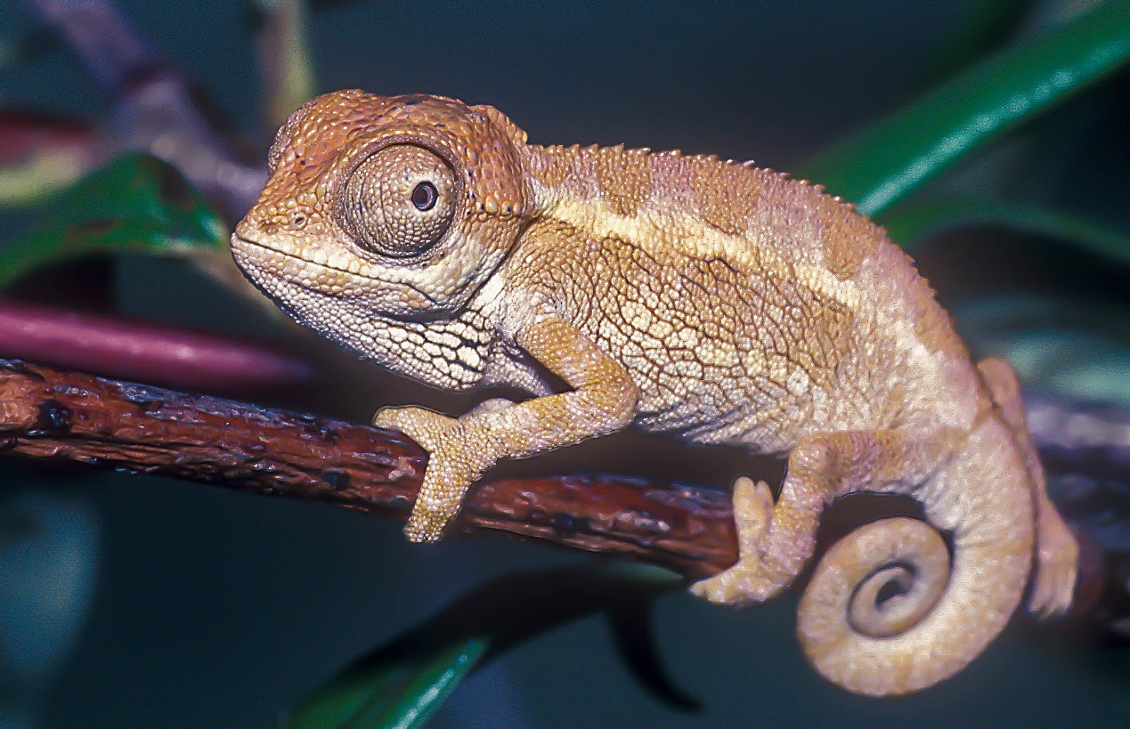
I was writing something on chameleons for another purpose when I wondered how many species I had kept and how many I had seen in the wild. That thought sent me into old records and trip reports. The first chameleon I kept was the small Two-lined Chameleon, now known as Trioceros bitaeniatus, from East Africa. In the 1950s it was stated that the ‘dwarf’ species lived longer than the larger ones. Quite by chance the way I kept it turned out to be in many ways ideal. Over two winters it lived on a wrought-iron framed mirror in the sitting room, eating flies from hatched fishing-bait maggots and mealworms held in forceps. In summer it was in a vivarium but on some days it was parked in an apple tree, with a piece of meat or fruit hanging by a thread to attract insects. At all times, the temperature fell at night—no central heating then. Later I had a single Jackson’s Chameleon (Trioceros jacksoni) and large number of Von Höhnel’s Chameleon, better known these days as the Helmeted or High-casqued Chameleon, Trioceros hoehnelii.


Natal Midland Dward Chameleon (Bradypodion thamnobates)
The late Bob Davies had a friend (whose name I cannot remember) who
started a colony of these chameleons in UK in the late 1980s.
I had some of the offspring which in turn bred.

Natal Midland Dwarf Chameleon - young
These chameleons bear live young

Natal Midland Dwarf Chameleon - a few days old


For decades attempts at keeping most species of chameleon in captivity for any length of time seemed doomed to failure. Indeed, they were one of the few groups of animals in which longevity was shorter in captivity than in the wild. However, that was only partly true since some species, for their size, do not live that long, some have very short lives, whereas others, the Veiled or Yemen Chameleon (C. calyptratus), for example, which proved to be easy to keep and breed will live for up to 5 (females) to 8 (males) years.

As with many reptiles, once captive-breeding was achieved with many species, subsequent breeding proved easier. The stress of capture, storage and transport was one reason for the lack of success. Better environmental conditions and nutrition also played a large part. I was astounded when I took some faecal samples from wild-caught chameleons in the 1980s. The parasite load was staggering: worms, protozoans and coccidial spores abounded. I could have sold seats by the microscope to those wanting to see a parasite zoo. It is not surprising that chameleons in general proved ‘difficult’ animals.

During the 1980s there was concern that since many species of chameleon survived so poorly in captivity that there would not exist reliable methods of ex-situ conservation should that prove necessary for some Madagascan species where habitat loss has been so dramatic. So many species have now been kept and bred successfully over a number of generations that such a concern has not been realised.


Carpet Chameleon (Furcifer lateralis)
This Madagascan species (sitting on the hand of the late Bob Davies)
is an egg-laying species. We both tried to breed this species but the
eggs were infertile. ca 1991

Flap-necked Chameleon (C. dilepis) ca 1991

Oustalet's Chameleon (Furcifer oustaleti)
Madagascar 2003


As much as I did learn about chameleons (and here I echo a former colleague who argued that if we learn how to keep and breed and animal in captivity then we know an awful lot—not everything but an awful lot—about how that animal works) there is nothing like seeing and observing animals in the wild. From Kenya in 1991, Madagascar (three weeks in 2003 and two days in 2006) and the Republic of Congo in 2014 we saw and watched chameleons in the wild. The fauna of Madagascar is of course amazing and for chameleon aficionado, who can see easily the radiation that has occurred there ranging from the tiny Brookesia to the huge Oustalet’s, one of the great wonders of the natural world.


Spectral Pygmy Chameleon (Rampholeon spectrum)
Republic of Congo 2014

And here are the lists I compiled, firstly of the ones I kept between 1959 and 1994:


Common NameScientific Name
Helmeted ChameleonTrioceros hoehnelii
Two‑lined ChameleonTrioceros bitaeniatus
Elliot's ChameleonTrioceros ellioti
Jackson's ChameleonTrioceros jacksoni
Johnston's ChameleonTrioceros johnstoni
Flap-necked ChameleonChamaeleo dilepis
Senegal ChameleonChamaeleo senegalensis
Carpet ChameleonFurcifer lateralis
Natal Midlands Dwarf ChameleonBradypodion thamnobates


...and secondly, chameleons I have seen in the wild from 1991 onwards:


Common NameScientific NameCountry
Stump-tailed ChameleonBrookesia superciliarisMadagascar
Spectral Pygmy ChameleonRampholeon spectrumRepublic of Congo
Short-horned ChameleonCalumma brevicorneMadagascar
Parson's ChameleonCalumma parsoniMadagascar
Nose-Horned ChameleonCalumma nasutumMadagascar
Malthe ChameleonCalumma maltheMadagascar
Oustalet's ChameleonFurcifer oustaletiMadagascar
Rhinoceros ChameleonFurcifer rhinoceratusMadagascar
Spiny ChameleonFurcifer verrucosusMadagascar
Jewel or Carpet ChameleonFurcifer lateralisMadagascar
Wills's or Canopy ChameleonFurcifer willsiiMadagascar
Panther ChameleonFurcifer pardalisMadagascar
Helmeted ChameleonTrioceros hoehneliiKenya
Flap-necked ChameleonChamaeleo dilepisKenya


Wednesday, 19 February 2020

A Hong Kong Sunbird fest

Hong Kong birdwatchers and photographers were out in force to see these 'Occasional Visitors' over Chinese New Year. Our Hong Kong correspondent was there, hiking from Shing Mun over Lead Mine Pass to Tai Po Kau.

Mrs Gould's Sunbirds (Aethopyga gouldiae) was what the birders were there to see. When first seen in Hong Kong they were regarded as escapees from captivity but it is now realised that Hong Kong is part of their natural range (from India, Nepal and Bhutan to Bangladesh, China,  Burma, Thailand, Laos and Viet Nam). First described in 1831 by the lawyer and politician Nicholas Vigors (1785-1840) while Secretary of the Zoological Society and pursuing his misguided 'quinarian system' of classification, it was named for Mrs Elizabeth Gould (née Coxon) (1804-1841) the artist wife of John Gould FRS, ornithologist, taxidermist and artist.


Mrs Gould's Sunbird - male


Mrs Gould's Sunbird - male. To show the iridescent feathers of the head


Feeding on the same flowers were 'Resident' Fork-tailed Sunbirds (Aethopyga christinae). Also called Mrs Swinhoe's Sunbird after the wife, Christina, née Lockie, of Robert Swinhoe FRS (1836-1877) who described the species in 1869, this bird in Hong Kong was regarded as a rarity when we lived in Hong Kong in the 1960s. First seen in Hong Kong in 1959, it could for years only be seen at Tai Po Kau. Since then it has spread throughout Hong Kong and on our first return to Hong Kong in 1997 a pair was nesting yards from our room at Robert Black College at the University of Hong Kong.


Fork-tailed Sunbird - male


Female sunbirds are easily overlooked:


Mrs Gould's Sunbird - female

And here are the photographers:


Also around was this Mountain Bulbul (Ixos mcclellandii) once a great rarity in Hong Kong but now regularly spotted:


Mountain Bulbul


A Verditer Flycatcher (Eumyias thalassinus)


Verditer Flycatcher - male


...and a female Red-flanked Bluetail (Tarsiger cyanurus) showing why it used to be called a bush-robin, a winter visitor to Hong Kong from the north:


Red-flanked Bluetail


Finally, a portrait of Mrs Elizabeth Gould with a pet Cockatiel. As well as illustrating John Gould's publication she had eight children, dying at the age of 39 after the birth of the eighth.


Sunday, 16 February 2020

Bouin’s fluid. But who was Bouin?

Bouin’s fluid will always be etched on my brain for a then commonplace but, with hindsight, foollhardy incident. During my final year at school I was a part-time laboratory steward (pay £2.14 shillings per week). One of my first jobs was to sort out the shelves at the back of the Advanced Biology Lab (now, along with the entire school, demolished). There was a 500 ml bottle half-full of Bouin’s fluid. The problem was that it had a ground-glass stopper. Even I knew that picric acid was an explosive and that anything containing picric acid should never come into contact with metal, must be kept dry and never put in anything with a ground-glass stopper. Friction or a sharp tap could detonate the dry material in the neck. I tried gently to remove the stopper. It was stuck. I had the wit not to try tapping the stopper on a bench to shift it because I could see dried picric acid in the neck. I then tried gentle heat from water heated with a bunsen burner (no piped hot water supply then) around the bottle. I grasped the stopper—there were no goggles or any other bits of safety equipment in the whole school—and pulled, without twisting. Out it came, but I don’t think I imagined hearing a small but definite ‘crack’. I now read that such episodes have to be dealt with by the bomb-disposal squad. I moved on to the next bottle. It was picric acid, obviously used in the past for the Bouin’s fluid. The contents were wet and it did have a rubber stopper.

Bouin's Fluid is available
ready-mixed
In the early and middle decades of the 20th century, the microscope was THE research tool in zoology—and many other -ology—laboratories. With the microscope came the paraphernalia to cut sections and stain the once-living material so that the various structures could be seen. Shelves in universities and schools were filled by a vast array of bottles containing various tissue fixatives, used to prevent shrinkage and decomposition, embedding materials, dyes, clearing agents, and mounting media for the thin sections. There was usually the evocative smell of Canada Balsam and xylol (then called xylene). Many of the mixtures of chemicals and dyes were named after the person who first used and described them during the golden age of histology. The obscure names on the faded labels of dusty bottles on equally dusty dark-stained wooden shelves added to the impression of alchemy rather than cutting-edge science.

Some of those named reagents have stood the test of time, while others have fallen by the wayside. The fixative of choice for the commonest staining method for mammalian tissues was and still is Bouin’s Fluid, a mixture of formaldehyde, acetic acid and picric acid. Having been aware of that fixative since the late 1950s, I had no idea who Bouin of his eponymous fluid was. Until last week that is when I saw a photograph taken at the First International Conference on Sex Hormones held in Paris in 1936. All of those present were well-known endocrinologists and I spotted those knew well, had met* or had heard of. And there was a Professor Pol Bouin of Strasbourg. Was he, I wondered, the Bouin of Bouin’s Fluid?


Pol Bouin circled
From Zuckerman's biography, From Apes to Warlords


Indeed he was. A bit of digging on Google soon turned up a biography. Pol André Bouin was born in Vendresse in northern France in 1870. While still a medical student in Nancy he became interested in morphology and was appointed preparateur d’histologie. He published the recipe of his new fixative in the same year that he qualified in medicine, 1897.

Pol Bouin
from Parkes
Bouin's rise in French science was meteoric, first in Nancy and then, after the First World War, in Strasbourg. His research covered a wide range of tissues and sub-cellular structures but he came to specialise in the reproductive organs and his fixative was devised for a study on seminiferous tubules. Much of his research and that of his collaborators in Strasbourg demonstrated the presence of putative hormones but preceded isolation and identification of the hormones themselves, prolactin and progesterone, for example. Similarly, his research in cytology, on the ergastoplasm (now the endoplasmic reticulum) was completely superseded once the electron microscope came on the scene.

Sir Alan Parkes—also on that photograph in Paris in 1936—wrote an obituary in Journal of Reproduction and Fertility. He began:

Pol Bouin, one of the pioneers of modern biology, and especially of the histological approach to physiological problems, died on February 5th, 1962, at the age of 92 at his family home in the Ardennes, to which he had retired finally in 1946. More than 20 years have elapsed since Bouin left active scientific work and time may have dimmed the bright light of his achievements for some of the younger generation. But in his own country, especially to his associates there, to the older generation elsewhere and to all with a sense of scientific history, Bouin ranks with F. H. A. Marshall and Ludwig Fraenkel as one of the small group of research workers who at the beginning of this century laid the foundations of our modern knowledge of the reproductive processes. Possibly even more significant than his personal original work was the influence he exerted directly and through his pupils. It may truly be said that Bouin founded a scientific dynasty which provided and continues to provide the major part of the great French contribution to our knowledge of the physiology of reproduction. 

But Parkes made no mention at all of Bouin’s fluid!


*Solly Zuckerman, Ruth Deanesly, Alan Parkes, Idwal Rowlands, Frank Young

Bouin first reported the use of his fixative in this paper:

Etude sur l'évolution normale et l'involution du tube seminifere. I. Modifications régressives du processus spermatogénétique provoquées expérimentalement. II. Phénomènes cytologiques anormaux dans l'histogenèse et l'atrophie expérimentale du tube seminifere. Arch. Anat. micr. 1, 225-265. 1897

Ortiz-Hidalgo C. 1992. Pol André Bouin, MD (1870-1962). Bouin’s fixative and other contributions to medicine. Archives of Pathology and Laboratory Medicine 116, 882-884.


Parkes AS. 1963. Pol Bouin 1870-1962. A memoir. Journal of Reproduction and Fertility 5, 301-307.

Monday, 10 February 2020

A Hong Kong Spider

Walking the hills last weekend, our Hong Kong correspondent spotted this large spider in a drainage ditch. We do not have a book on Hong Kong spiders but it looks like the huntsman, Heteropoda venatoria which goes by a number of common names including Giant Crab Spider. Huntsman spiders are straight killers, building no web to trap their prey. The venom contains a potassium channel-blocker—bad news for anything with a nervous system. Insects are its main prey but it has, apparently, also been known to kill scorpions and bats.