Sunday 27 June 2021

Chinese Mountain Cat: New research indicates it is a subspecies of the Wildcat

After our encounters with Chinese Mountain Cats in Sichuan in 2017, I wrote several articles on this enigmatic and attractive small felid (here, here, here). Over the years, the origins and status of this cat have been discussed. In essence, the argument has been whether it is a full species, Felis bieti, or a subspecies of the Wildcat, here called Felis silvestris. At the time I wrote those articles the view that appeared to prevail was that since it had been reported that the ranges of the Chinese Mountain Cat and the Wildcat overlap, i.e. they are sympatric, they must, therefore, be two different species.


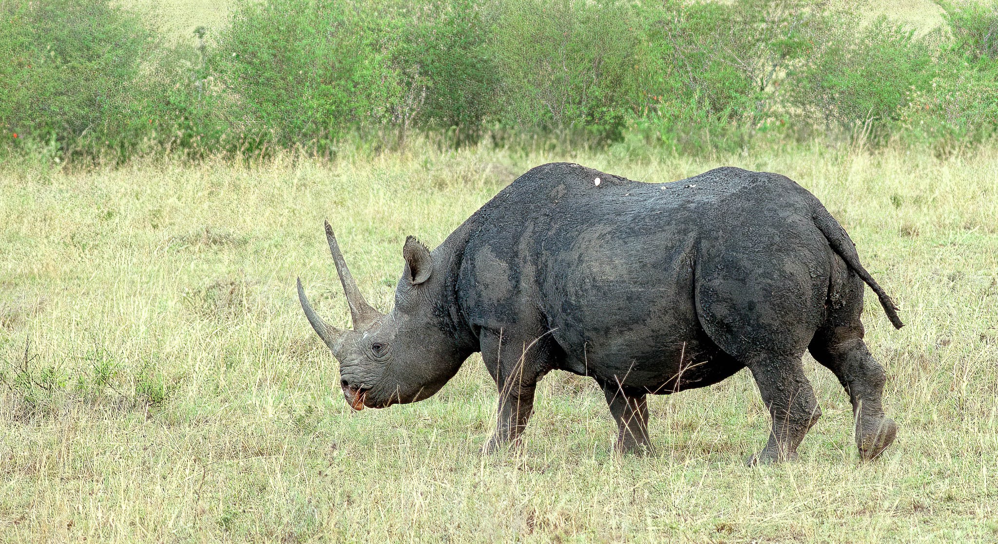
These photographs of a Chinese Mountain Cat were taken by Tim Melling
in 2019 in the same location we (with Tim) saw them in 2017.
This one was crouched next to the road and he took this photograph
from the car using torchlight in the pitch dark. The light blue eyes
and the ear tufts can be seen. Note the thick, unpointed bushy tail.
I wonder if this is the same cat as the one I videod at long range
as it patrolled the grassland in 2017?
These are from Tim Melling's Flickr pages here


The Chinese Mountain Cat was first described by the Frenchman of British descent and English surname, Alphonse Milne-Edwards, in 1892. At the time Milne-Edwards was the new Director of the Muséum National d'Histoire Naturelle in Paris. He named the cat, collected in Sichuan, as a new species, Felis bieti, after the French missionary and naturalist, Félix Biet (1838-1901).


Before describing the latest research I must point out that the taxonomy of the various forms of Wildcat in Eurasia and Africa has been the subject of constant change. I am following the authors of the current paper in calling the Wildcat of Europe and parts of Asia, Felis silvestris with five distinct interfertile subspecies or geographical forms, the one in northern China being Felis silvestris ornata. An alternative scheme from 2017 lumped the Wildcats from Africa and Asia into one species, Felis lybica; in that scheme the Wildcat of northern China is Felis lybica ornata. However, that wider problem of Wildcat classification is not germane to the present research on the status of the Chinese Mountain Cat and its relation to the northern Chinese form of the Wildcat.


This week a new paper appeared in Science Advances which specifically addresses the status of the Chinese Mountain Cat. A group of authors from China, Malaysia, Russia and the USA have done a phylogenetic analysis of zoo, museum and villages-kept specimens using nuclear and mitochondrial DNA sequences of 27 Chinese Mountain Cats, 4 Wildcats of the form found in China (Felis silvestris ornata) and 239 domestic cats.


From the phylogenetic analysis, the authors conclude that the Chinese Mountain Cat, which occurs only on the Qinghai-Tibet Plateau, is not a separate species but a subspecies of the Wildcat with a past and complex history of hybridization between the two. Surprise, surprise: Mountain tom cats spreading their genes into the Wildcat population and the offspring back-crossing into the Mountain population appears the most likely explanation for the hybridization events at a time when the ranges of the two forms overlapped. Evidence of extensive past genetic exchange between the two lineages and therefore interbreeding was found


The authors also address the question of whether this conclusion from nuclear and mitochondrial DNA is also compatible with ‘the biological species concept, which considers interbreeding as the prerequisite for a species. The key argument from the proponents for the species status of the Chinese mountain cat lies on its distinctive morphological characters, a presumed sympatric distribution with the Asiatic wildcat, and an absence of gene flow between free-ranging Chinese mountain cats and Asiatic wildcats. However, recent surveys in Northwest China showed that the range attributed to the Asiatic wildcat may have been overestimated and that its presumed presence on the Qinghai-Tibet plateau in north-eastern Qinghai may not be true. That assertion, if proven, would dispute the supposed sympatry of the two lineages’


Differences in appearances were noted in genetically-determined hybrids. For example, the tail was less bushy in a cat with the nuclear DNA of a Mountain Cat and the mitochondrial DNA signature (passed down the maternal line) of a Wildcat. The presence of hybrid cats (i.e. crosses with Wildcat or with domestic cat—see below) could explain the seemingly anomalous appearance of some cats seen and photographed on the Qinghai-Tibet Plateau in recent years.


The authors continued in their conclusion on the Mountain Cat:


Answers to the remaining questions require more surveys and studies to fine map the Asiatic wildcat and Chinese mountain cat distribution in Northwest China; to delineate the subspecies boundaries or hybrid zones; to elucidate the ancestry, adaptation, and evolution of these taxa; and to resolve the historical and current patterns of gene flow among the wildcat and domestic cat lineages in the region.


Will the status and origins of the Chinese Mountain Cat and the other Wildcats of Eurasia and Africa now be regarded as settled? I wouldn’t bet on it although at the moment the ‘lumpers’ certainly hold sway.


The point the authors make on evidence of recent cross-breeding with domestic cats has major implications for the conservation of the Mountain Cat, as the authors explain:


Contemporary genetic introgression from F. s. bieti into sympatric domestic cats is evident across, but not beyond, the range of F. s. bieti. The timing of admixture coincided with large-scale socioeconomic changes in the Tibetan area during the mid-20th century. That process likely led to an expansion of domestic cats into the region and suggests that domestic cats arrived rather late to the Plateau and thus had not encountered F. s. bieti until recently. The increasingly abundant local domestic cat population may pose a threat to the Chinese mountain cat and jeopardize its genetic integrity and evolutionary adaptation to high altitude, an issue with profound conservation implications and worth further study.


Those of us who live in Scotland will recognise the same conservation and taxonomic problems with the Scottish form of the Wildcat. Firstly, survival in the wild is severely threatened by cross-breeding with domestic cats. Secondly, the status of the form has varied from being considered a species (Felis grampia) a separate subspecies of the Wildcat (F. silvestris grampia) or lumped into the European Wildcat (Felis silvestris silvestris). I read that the latter is the currently favoured view.


On that note of deep concern for the future integrity of the Chinese Mountain Cat, the present data supporting ‘lumping’ of the Felis cats mean China loses an endemic species from its faunal list but gains yet another conservation problem.


He Yu, Yue-Ting Xing, Hao Meng, Bing He, Wen-Jing Li, Xin-Zhang Qi, Jian-You Zhao, Yan Zhuang, Xiao Xu, Nobuyuki Yamaguchi, Carlos A Driscoll, Stephen J O’Brien, Shu-Jin Luo. 2021. 

Genomic evidence for the Chinese mountain cat as a wildcat conspecific (Felis silvestris bieti) and 

its introgression to domestic cats. Science Advances 7 (26), eabg0221. DOI:10.1126sciadv.abg0221 


Thursday 24 June 2021

The Reptile House at London Zoo: the beginning of the end

News that London Zoo was to start the process of planning a new reptile house appears to have evoked little response other than in the zoo enthusiast community which, in general, has expressed regret at the loss of the old one. The plan, as reported, is for the new one to hold only 29 species—all for captive breeding of species of conservation concern, plus a new house for Galapagos tortoises, while the old house is to be retained for purposes yet to be decided.

Views of the current Reptile House have varied over the decades from its opening in 1927. It was groundbreaking in terms of design, control of environmental temperatures and provision of ultraviolet radiation but a common view in the 1950s and 60s was that it was a deathtrap for its reptilian inhabitants (it had soon been established that it was too hot for many amphibians). Design of the house was attributed by the besotted Peter Chalmers Mitchell to Joan Procter with the architect Guy Dawber being demoted in importance to having added the fancy bits. By contrast, Solly Zuckerman thought Joan Procter overbearing, over-rated and over-promoted.


Entrance to the Reptile House, 2011
Photograph by William Hook via Wikimedia

Original plan of the Reptile House
The Times 15 June 1927

It is true that the mortality rate of reptiles at London Zoo was very high in the early decades of the reptile house’s existence. in 1946-48 the annual death rate was 45.4%; in 1956 it was 68%. In other words two-thirds of the inhabitants were dying within the year. I do not know the extent to which the environments afforded by the house were responsible. However, it is worth pointing out that many reptiles and amphibians arrived from abroad both for zoos and animal dealers in very poor condition. Animals were often unfed and unwatered for long periods and kept in crowded, unsuitable conditions before being shipped. Reptiles take a long time to die and can reach a stage of irreversible metabolic damage. No matter how well they were treated on arrival the outcome was inevitable. The great advances in reptile husbandry (and in the veterinary treatment of wild animals) of the 1970s and 80s was still to come. From being unable to breed nearly all species in the 1950s, private herpetologists led the way in devising the means of doing so. Zoos generally followed that lead but it was clear that the Reptile House was not ideal for use as a breeding setup even with the best efforts of the keepers at the time. Furthermore, the building was getting tired with much of the old equipment unusable; for long periods of its existence there was not enough money available for even minor improvements. Even 20-30 years ago it was evident to many of us that the Reptile House needed to be replaced in order to provide accommodation commensurate with the great advances made in keeping reptiles during the 1970s and 80s and with providing staff with better facilities for incubation of eggs and the raising of young animals to adulthood together with showing visitors these processes in action.

I am therefore delighted to hear the plans for a new reptile house. I do though share the concern of comments I have read that plans are being made for only 29 species. That announcement cuts to the heart of the matter about the role of zoos in the 21st century. Should the collection comprise only those species in actual or potential need of captive-breeding programmes? Or should they be there to educate the public about the diversity of animal species? How much public ‘entertainment’ in the way of common but popular species or ‘immersive’ exhibits is it necessary to include in order to attract the paying public in sufficient numbers and thereby support financially the conservation and educational efforts within the zoo and in the wider world?

These strategic questions and the arguments for and against each scenario are not new and will continue to engage informed—and much uninformed—opinion. On the ground it is difficult for the majority of zoo visitors to appreciate how many of what were major collections, including London Zoo, have destocked in terms of number of species and of individual animals kept. Keeping the discussion to reptiles and amphibians, in the years 1949-51 London Zoo had 221 species; in 1957 there were 168 while in 2020 the number was 59.

What concerns many people I know is the lack of a comprehensive collection of reptiles and amphibians in Britain. With so few native species, these animals are the least known and, despite decades of educational effort, the most reviled. They need to be seen in the flesh in all their diversity. Television just does not cut it. There is a real opportunity for imaginative displays, some with ‘reversed daylight’ to show the diversity, adaptations and breeding of reptiles and amphibians and why they are an important part of the natural world.

The days though of going to the Reptile House first to see what was happening and what was new—a common trait amongst many of us whose first interest was reptiles and amphibians—seem numbered.


Tuesday 22 June 2021

Masai Mara Safari - 30 years ago

Itchy feet with the world out of reach? These photographs from the Masai Mara in September 1991 will not help.


Black Rhinoceros

Topi (Damaliscus lunatus) with Thomson's Gazelles (Eudorcas thomsonii) on the right.
Landscape typical of the Masai Mara

Lioness

Cheetah

...only moved slightly when we passed by later


Friday 18 June 2021

Marsupial frogs: Important questions remain on life in the pouch

Following up my article on marsupial frogs, I have acquired a copy of William Duellman’s book, Marsupial Frogs. Gastrotheca and Allied Genera published in 20151. Book hardly does the work justice because it a complete account, based on five decades of research by the author, particularly on their taxonomy and evolution, of this remarkable group of South American amphibians. As well as general chapters on aspects of their biology, each species is described in detail. Duellman began his book as follows:

What are marsupial frogs, and why are they so particularly worthy of study. These are the only frogs in which males fertilize the eggs out of the water and then place them in a pouch on the back of the female. The developing embryos of marsupial frogs and their allies—i.e. the amphibian family Hemiphractidae—have large external bell-shaped or sheet-like gills that are unlike those of any other lineage of frogs. These behavioral and developmental features are unique to marsupial frogs and their allies.


The first point I wanted to check was what he had to say about the identification of frogs exported from South America in the 1950s, presumably through the animal trade. These animals were the basis of early work on reproductive behaviour and the use of the dorsal sac to hold fertilized eggs. As I stated in my previous article, the frogs are now thought not to have been Gastrotheca marsupiata or marsupiatum as they were labelled at the time, but G. riobambae from Ecuador. Duellman in his book repeats the sorting out of this misidentification but also adds that another species, G. pseustes, very similar in appearance to G. riobambae, also occurs in the Andes surrounding Quito; that species may have been also have been involved in the exports and studies.


Getting the species right is important in the these frogs (69 species recognised in the genus Gastrotheca at the time Duellman’s book was written) because the stage of development at which the young emerge from the pouch on the mother’s back differs. In a few species, incidentally, the pouch is on the side and even extends via a slit into the abdominal cavity. Some hemiphractid species, all in genera other than Gastrotheca, have no fully enclosed pouch: some have a dorsal patch to which eggs adhere; others have a basin-like structure while in one genus, the eggs are enclosed but the two edges of the skin flaps forming the cover do not fuse.


From Duellman's book


In species such G. riobambae, G. marsupiata and G. pseustes the tadpoles when they hatch and shed their external gills leave the pouch (assisted by the toes of the female being inserted into the pouch) and continue their feeding, development and metamorphosis in ponds. The tadpoles, it should be noted, hatch at a more advanced stage than in species like the Common Frog. By contrast in some other species of Gastrotheca, the young hatch and emerge as fully-developed froglets; metamorphosis takes place in the egg capsule within the mother.


This is where things get physiologically more interesting. Marsupial frog tadpoles develop large external gills connected to the tadpoles by stalks. The gills have a large surface area and form, by analogy with mammals, the fetal side of the placenta. The brood pouch is formed from skin but internally loses loses much of the structure typical of skin when eggs are in the pouch. The epidermis becomes thin and produces well vascularized extensions that partially envelop the egg. In other words, the skin forms the analogue of the maternal side of the mammalian placenta. Between the two sides is the egg capsule across which any exchange or transfer of materials between mother and egg/tadpole must occur. It is obvious that oxygen must pass from mother to egg/tadpole with carbon dioxide moving in the reverse direction. The form of the nitrogenous waste produced by the tadpoles shows an interesting adaptation. Free-living tadpoles usually resemble fish in that they produce ammonia—they are ammonotelic—which being toxic is swiftly excreted into the surrounding water; at metamorphosis terrestrial amphibians switch to produce urea. However, marsupial frog tadpoles produce urea—they are ureotelic—and not ammonia. Not all the urea though leaves the egg capsule. It has been suggested that the urea could serve some osmotic function (as in the Crab-eating Frog) and preventing the loss of water. This aspect required further study but the inclusion of urea in the culture medium made it possible to keep early embryos of G. riobambae alive after they were removed from the pouch; the usual saline solution was not sufficient.


Much of this work on the developmental biology of marsupial frogs has been done by Eugenia del Pino in Quito2; she has worked extensively on G. riobambae, and the aspects of her research described here only skim the surface of her studies done over the past 50 years on these fascinating frogs and their adaptations. Her research as a developmental biologist therefore complements that of Duellman, centred on evolution and taxonomy, over a similar period of time.


The big question, of course, with marsupial frogs is whether or not the developing tadpoles draw on maternal nutrients to support their growth and development in addition to the nutrients provided at the time of laying in the yolk—the eggs of marsupial frogs are very large and therefore contain a lot of yolk. In the tadpole-producing G. riobombae there is evidence that the developing tadpole relies solely on the yolk until it hatches and leaves the pouch. The weight of dry matter in the egg capsule was found not increase during development. By contrast, a more recent study3 on a froglet-producing species, G. excubitor, has suggested that maternal nutrients could be transferred to the developing young. However, having read the paper and having looked at the supplied raw data I remain unconvinced. The number of animals studied was very small at each stage of development; the embryos were only studied at early stages of development and the results of feeding stable carbon and nitrogen isotopes to prey insects, while indicating that carbon and nitrogen compounds (which could be simple products of metabolism like carbon dioxide or urea) pass from mother to embryo through the egg capsule, are not a demonstration of NET transfer of nutrients.


There do appear to be adaptations associated with producing froglets rather than tadpoles. Duellman provided a diagram showing that froglet-producing species have fewer, larger eggs (thereby providing evidence that the yolk is sufficient for development to froglet); eggs in the pouch are arranged in a single layer such that they can be completely enveloped in maternal blood vessels from both sides. By contrast, tadpole producers have more, smaller eggs, stored as a double layer in the pouch. All these findings are consonant with the increased requirements for oxygen in the later stages of growth and development by the larger tadpoles and metamorphs of the froglet producers.


In conclusion, the question remains—as it does for other frogs in which eggs are held in various body cavities until newly-metamorphosed froglets emerge—do maternal nutrients contribute to the metabolism and growth of the developing tadpole? The simplest way of answering that question would be to weigh the dry matter of the egg capsule and its contents right through the period of development in the pouch, as has been done in the tadpole-producing G. riobambae. We would then know if then know if the egg capsule with its rich supply of blood vessels on both sides acts in a manner more comparable with the mammalian placenta or if it just, as it says on the tin, just a gill. Molecular biologists might want to just have a look at whether or not transporters for glucose and amino acids occur in the egg capsule. 


The question of whether the external gill acts as a gill or an analogue of the mammalian placenta takes me full circle to my start of the first article and why Amo was fascinated by marsupial frogs and had a film made of their reproductive behaviour. There is still much of interest to do, with key questions still waiting to be answered, 65 years later.


1. Duellman WE. 2015. Marsupial Frogs. Gastrotheca & Allied Genera. Baltimore: Johns Hopkins University Press.


2. del Pino, EM. 2018. The extraordinary biology and development of marsupial frogs (Hemiphractidae) in comparison with fish, mammals, birds, amphibians and other animals. Mechanisms of Development 154, 2-11.


3. Ware RW, Catenazzi A. 2016. Pouch brooding marsupial frogs transfer nutrients to developing embryos. Biology Letters 12: 20160673. http://dx.doi.org/10.1098/rsbl.2016.0673 


Sunday 6 June 2021

‘A quantity of rhinoceros (defunct) on the premises’: the discovery of the discoverer of the parathyroid glands

There are not many articles that can be started with the immortal line of W.S Gilbert ‘When I was a lad’. But this one can.

When I was a lad the discovery of the parathyroid glands was attributed to the Swedish anatomist, Ivar Sandström (1852-1859), of the University of Uppsala. He described these ‘new’ organs closely attached to the thyroid in man and the separate organs in the horse, rabbit and cat. Their vital function in calcium metabolism was of course unknown—Sandström thought them to be embryonic portions of the thyroid—and the name ‘parathyroid’ only came into use in 1896.

It was only gradually in the second half of the 20th century that historical discoveries published in 1953 became better known. The outcome, in short, was the recognition of Richard Owen, that great anatomist, anti-Darwinian and deceitful oleaginous creep, was the first to describe the parathyroids. Now, in contrast to 60 years ago, Owen and not Sandström appears in all the literature as the discoverer

The possibility that Sandström may not have been first was raised by the physician, Sir Humphry Davy Rolleston (1862–1944) in a book on the endocrine glands published in 1936: 

The presence of what were, or may have been, parathyroids was observed by Robert Remak (1815-1865) of Berlin in 1855, by Richard Owen (1804-1892)…in 1862 in an Indian rhinoceros…and by [R.] Virchow (1821-1902) in 1863 in man.

That extract was from a chapter of a book published in 1953 and written by Alexander James Edward Cave (1900-2001). I will return to Professor Cave, who uncovered the whole story of the discovery of the parathyroids, below.

Cave established that Owen had indeed described what was later named the parathyroid glands but even so, the date given by Rolleston, 1862, for Owen’s paper would still have left Remak the probable first—in 1855. Cave, though established that Rolleston had misinterpreted the date of Owen’s publication in Transactions of the Zoological Society of London. 1862 was the date the whole volume was published but each part of the volume had previously been published separately. He explained:

The work of dissection and drawing proceeded apace during the winter months of 1849-50, and on 12 February 1850 Owen communicated the resultant monograph to a meeting of the Zoological Society. This paper was published as the third article in the fourth volume of the Society's Transactions, which volume, covering the period January 1851 to September 1862, bears the terminal date of 1862—a point which has led Rolleston and possibly others astray. For in those early days of the Zoological Society's publications, the individual papers comprising any particular volume of its Transactions appeared separately, under dates anterior to the terminal volume date. Thus Owen's rhinocerotine memoir was published, not in 1862 as commonly supposed, but full ten years earlier, viz., on 2 March 1852, a point of importance in connexion with priority in the matter of the discovery of the parathyroid glands. 

Owen really had got there first and in a rhinoceros, the Indian species Rhinoceros unicornis, which had died at London Zoo.


Richard Owen discovered the parathyroid gland in an Indian Rhinoceros
We saw this mother and offspring Kaziranga in Assam in February 2007


Cave himself had produced a series of papers on the anatomy of rhinoceroses which, again, like Owen, based on the dissection of animals that had died at London Zoo. He knew what he was looking for when he re-examined Owen’s descriptions. The clincher was the finding that Owen’s dissection of the thyroid region had been preserved and had survived in the museum of the Royal College of Surgeons. Cave himself had dissected the same region in the neck of two Indian rhinos that had died at London Zoo in 1941 and 1945. He knew what he was looking for and found the dissection ‘tedious and laborious’. He was full of admiration for Owen, dissecting in unknown territory but uncovering and displaying an organ ‘in diameter and circumference no greater than a sixpenny piece’ (i.e ¾ inch or 19.3 mm diameter). 


Cave included a photograph of Owen's dissection preserved
in the museum at the Royal College of Surgeons


Owen was dissecting the first Indian Rhinoceros to be exhibited at London Zoo. He described the animal as his ‘ponderous and respectable old friend and client’, client because he had examined the animal before its purchase by the zoo fifteen years earlier ‘and took it upon my skill, in discerning through a pretty thick hide the internal constitution, to aver that the beast would live to be a credit to the Zoological Gardens, and that he was worth the 1000 guineas demanded for him’. In present day values the price was around £100,000.

After it death on 19 November 1849 dissection of the beast, which would have weighed over 2000 kg, was done at the Royal College of Surgeons where Owen, as Conservator of the museum, had a residence. Owen’s wife, Caroline, recorded in her diary that 'as a natural consequence' of this animal's death 'there is a quantity of rhinoceros (defunct) on the premises’. She was probably inured to such matters; her father was the first conservator of the museum. And inured she probably needed to be since the rhinoceros flesh would have been putrefying after several months in the building. Although the retained specimens would have been preserved in spirit, the use of formalin for temporary preservation (as in our dogfish, frog and rabbit dissection days for ‘A’ levels) did not arrive until nearly the end of the 19th century. The only consolation for Mrs Owen is that the animal died in November and not June.

Cave ended his chapter:

That Owen was the first to describe and to preserve the gland now called parathyroid, and that he recognized its glandular nature, is sufficiently established. His discovery deserves to be more wideIy known and appreciated, not only as constituting a tribute to his acumen as an investigator, but also as redounding to the credit of British anatomical science. 

The term 'parathyroid' is admittedly unsatisfactory as a descriptive label for structures which, in different mammalian species, and even in different individuals of any one species, may be indiscriminately para-, epi- or intrathyroid in position. For want of a more convenient term, and by general acceptance, this name is established and is likely to remain. At one period, however, the parathyroids were commonly known as 'Gley's glands'. Eponymous anatomical nomenclature is, unfortunately, unfashionable nowadays—to the detriment of the student's appreciation of the long, varied and educative history of the subject. But should such nomenclature achieve a return to favour, the parathyroids are already provided with their most appropriate name—‘the glands of Owen’.

Before ending this article on an endocrine gland in a ponderous pachydermatous perissodactyl it would be remiss to omit more mention of the man who discovered the discoverer, Professor Cave.

Professor Cave

A.J.E. Cave

Professor Alexander Cave was possibly the last surviving classical anatomist in Britain. Like some of his contemporaries he did not confine himself to human anatomy and in his later years made the anatomy of rhinoceroses his particular interest. He worked in London and from London and Whipsnade zoos he had over many decades a number of animals to study. However, he also sought out preserved specimens in museums and studied some aspect of the anatomy of all species. His papers on rhinoceroses can be found in full at the Rhino Research Center’s website. However, he by no means confined himself to rhinos; he wrote papers on a multiplicity of topics and species, including whales and gorillas, neanderthals and human acrobats. His final paper was published at the age of 94.

Cave was, being an anatomist then, medically qualified. From being a medical student in Manchester he moved, first to Leeds for 10 years and then to University College London. In 1935 became Assistant Conservator of the Hunterian Museum of the Royal College of Surgeons, and from 1941 held the title of Professor of Human and Comparative Anatomy. He was in post when, at 12.55 am on 11 May 1941, bombs hit the building and destroyed half of the collection of specimens. In 1946, Cave was appointed to St Batholomew’s Hospital medical school as Professor of Anatomy. There he stayed until he ‘retired’ in 1967. Students who passed through his lecture theatre, dissecting room and examination halls found him terrifying—a common trait of professors of anatomy at a time when the subject dominated preclinical medical education—but also a highly-regarded supportive figure. He is said though to have found difficulty coming to terms with the presence of increasing number of female students. After his retirement he moved his base to London Zoo where be became a willing horse, editing papers for ZSL’s scientific journals and chairing innumerable scientific meetings.

As well as wide-ranging interests in things conventionally anatomical, Cave had a deep interest in the history of the subject—as is evident from his work on ‘the glands of Owen’. He, a staunch Catholic, was also called upon to determine the authenticity of relics in various religious establishments, both Catholic and Anglican. In a report never made public or retained (except for the one copy kept by Cave) he found no evidence to support the belief that bones in a shallow grave in Canterbury Cathedral were those of Thomas à Beckett murdered in 1170. That must have come as a shock to the Dean and Chapter; they had already commissioned an architect to design a cover for the burial place. In other cases he variously decided for (the skull of St Ambrose Barlow, who was hanged, drawn and quartered at Lancaster Castle) and against (the alleged hand of St James the Great at a church in Buckinghamshire) the authenticity of other relics.

Very little was known about Professor Cave, where he came from and what he did, by younger attendees of scientific meetings at ZSL in the 1970s. He appeared, chaired the meeting with avuncular charm and sometimes misunderstanding—and then disappeared. One obituarist remarked that having been born in 1900, “he was a Victorian, and something of that period clung to him: in his dress, in his splendid use of the English language, and, yes, his gentle gallantry with the ladies”.

Having been a lay brother in a monastic order, on his second wife’s death he moved into a convent care home; there he celebrated his 100th birthday eight months before his death.

Finally, Cave complained to his opposite number at the Middlesex Hospital that his office at Barts was rather small. There was a swift rejoinder to the effect that any office would look small if it was, like Cave's, shared with the skulls of two adult rhinoceroses.


Cave studied all the rhinoceros specimens he could lay his hands on
This is a Black Rhinoceros we photographed in the Masai Mara in September 1991


Cave AJE. 1953. Richard Owen and the discovery of the parathyroid glands. In, Science Medicine and History Volume II. Edited by E Ashworth Underwood, p 217-222. London: Oxford University Press.

Cave AJE. 1953. The glands of Owen. St Bartholomew’s Hospital Journal 57, 131-133.

Cave AJE. 1976. The thyroid and parathyroid glands in the Rhinocerotidae. Journal of Zoology 178, 413-442.

Walls E. 2001. Professor A.J.E. Cave 1900-2001. Journal of Zoology 255, 283-284.