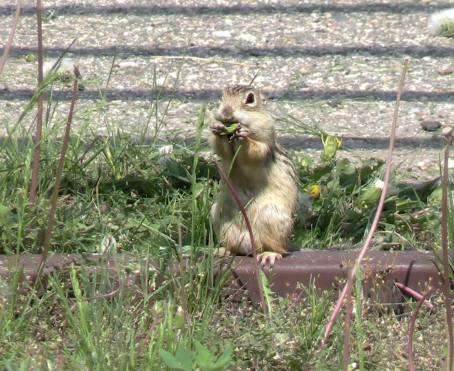
In a previous article I described my delight at seeing Thirteen-lined Ground Squirrels for the first time in the wild. These animals are a common sight in the grasslands and prairies of north America. My first encounter with this species was 58 years earlier. Before I joined him as a PhD student John Phillips (1933-1987) was back in Sheffield on his first long leave from the University of Hong Kong where he was Professor of Zoology, having been appointed three years earlier at the age of 29. In those days expat terms were such that after two years and seven months ‘home’ leave of five months was taken—an ideal arrangement for those wishing to do research. I found him in the attic, used as an animal house. He was concerned that a group of ground squirrels - kept together—was not thriving. I could see they were being fed on standard rat lab pellets plus the usual mixture of seeds, biscuit etc given to hamsters. I immediately suggested giving them more protein since I had kept a related species and knew that they ate insects and other invertebrates in addition to the typically rodent diet. The technician in charge could not believe I was right but went off to collect a handful of mealworms which were thrown into the floor cage. The ground squirrels fell upon them and had soon scoffed the lot. With the problem of feeding solved, John finished his work on them and returned to Hong Kong. I assumed that the work had been published since in his report to the Nuffield Foundation in 1967, which funded his work in comparative endocrinology to the extent of pay, equipment and consumables for a research unit in Hong Kong, he listed among the publications one on the Thirteen-lined Ground Squirrel which was in preparation for submission to the
Journal of Endocrinology. However, when I searched all his publications while researching this article I could not find it and only this week have I found that the work was never published and realised why it was never published
In the early decades of the 20th century, endocrinology was in its infancy and there was still argument, dating from the 1850s, of whether or not the adrenal glands are essential for life. The Thirteen-lined Ground Squirrel (now Ictidomys tridecemlineatus but then included in the genus Citellus and later Spermophilus) achieved its fame as an oddity in comparative endocrinology because it can consistently survive removal of the adrenal glands by virtue of the growth of adrenocortical tissue in the ovary. Richard Arnold Groat (1915-1994) was a PhD student in the Department of Anatomy at the University of Wisconsin when he published his findings in 1943 and 1944. Groat went on to work in a number of medical schools and in private practice as a pathologist in North Carolina.
Groat showed that in common with a number of other species, Thirteen-lined Ground Squirrels only survive after adrenalectomy if given 1% sodium chloride to drink. With that treatment the animals continue to feed. However, unlike in other species, Groat found that the salt water can be removed after a minimum of 15 days and that the ground squirrels can withstand a fast after 3-4 months. He then showed that adrenocortical-like tissue appeared in the ovaries after complete removal of the adrenal glands strongly indicating that adrenal steroid hormones were being produced by the ovary in quantities sufficient for normal life.
I do not know how Ian Chester Jones (1916-1996) heard of Groat’s work. There was no mention of it in his seminal monograph on the adrenal cortex which was published in 1957. However, he became sufficiently interested to start this work in 1961 at Harvard. He was again working as a visitor in Roy Orval Greep’s (1905-1997); he had spent a couple of years there in the 1950s on a Fellowship of the Commonwealth Fund of New York (later to become Harkness Fellowships). Ian Henderson, then a PhD student in Sheffield (1941-2019) was the second author of the paper published in 1963. They explained their interest in Groat’s work:
His [Groat’s] findings have always been open to the suspicion that they are merely manifestations of adrenal rests which showed particular responsiveness in this species. On the other hand, his work may indicate the capacity of ovarian tissue to change into a new type which may be adrenocortical both in form and function. Should this be so, then a question of fundamental importance is raised. It may be, for example, that the ground squirrel displays, in intensified fashion, a property that exists in mammals generally. For these reasons, this paper presents a re-examination of the problem.
In modern terms, there is another possible explanation: a population of pluripotent stem cells or partially differentiated cells in the ovary stimulated by ACTH from the pituitary to divide and differentiate into cells typical of the adrenal cortex rather than of the ovary, the ovary and adrenal cortex being of similar embryonic derivation. With the negative feedback between concentrations of adrenal steroid hormones and ACTH secretion, the concentrations of ACTH in the blood would have been sky high in the adrenalectomized animals.
Essentially Chester Jones & Henderson confirmed Groat’s findings. As with Groat, they were unable to find adrenal rests—small isolated clumps of adrenocortical tissue outwith the adrenal gland—in the normal adrenal. These occur in various locations including the ovary and testis in mammals.
The implication of the observations would be that with the apparent absence of adrenal rests, the ovary of the intact ground squirrel would not be producing adrenal steroid hormones. However, Gavin Vinson (1939-2021) then also working with Chester Jones in Sheffield found that ovarian tissue could convert progesterone to the adrenal steroid hormones, corticosterone. In his paper of 1965 he wrote (references omitted):
It is particularly interesting that the ovary has the capacity to convert progesterone to corticosterone. While this might be ascribed to adrenal rests, Groat (1943, 1944) and Chester Jones & Henderson (1963) were unable to find adrenocortical tissue in the ovaries and adnexia of normal ground squirrels. Another possibility is that ovarian cells, possibly the interstitium, of the ground squirrel are able to produce both sex and adrenocortical steroids. An attractive hypothesis is that, as the ovary shares a common embryological origin with the adrenal cortex, throughout the vertebrates both tissues are able (to a greater or lesser degree) to manufacture both oestrogens and corticosteroids.
While there is current molecular evidence against the latter in the latter explanation in mammals, I suspect the explanation possibly lies in a small population of partially differentiated adrenocortical cells in the ovary of the ground squirrel, i.e. cryptic and widely spread adrenal rests with the potential to grow into clumps of adrenocortical tissue.
William George Henry Seliger (1922-2016), A James Blair and Harland Winfield Mossman (1898-1991) followed up Groat’s work in 1966; they were in the same department as Groat in the University of Wisconsin and I suspect Groat may have been Mossman's student. They showed that the cells characteristic of the adrenal cortex which appear in the ovary are in close relationship to the epithelial remnants of the rete ovarii and the medullary cords. Identical cells were found around the efferent ductules of the testis in adrenalectomized males. Histochemical tests indicated the cells were identical to normal adrenocortical cells. In vitro, the areas of the ovary with the adrenal-like cells converted progesterone to adrenal steroid hormones identified as corticosterone and desoxycorticosterone (11-deoxycorticosterone)* the combination providing both glucocorticoid and mineralocorticoid activity. Confirmation of the vital role of the ovaries and testes in adrenalectomized ground squirrels was obtained:
Male and female animals adrenalectomized a year before and maintained on tap water died within four days when the gonads and adnexa were removed. Male animals adrenalectomized a year before and maintained on tap water survived actively for two weeks when the testicles were removed, provided that the efferent ductules and surrounding adrenal cortex-like cells were left in place. These animals died within four days after the adnexa were removed.
To return to the paper by Phillips and Chester Jones that was never published, the chronology suggests that the work they were doing in September/October 1965 produced results indistinguishable from those published by Seliger, Blair and Mossman in 1966. In short, they had been scooped.
Since the 1960s there has been little to add to the story. It would seem that cells with the potential to become adrenal cortical cells can be retained during embryonic development in the ovaries and testes in the ground squirrel. In that respect there is nothing new—adrenal tissue has been found after adrenalectomy in similar locations of mesodermal origin in other mammals. In the rat a report from the 1920s indicated an incidence of 19%. Similarly, some human patients show accessory adrenal tissue after adrenalectomy. However, what makes the Thirteen-lined Ground Squirrel special is the extent and consistency of formation of adrenocortical tissue in the ovary and testis. The finding in 1967 of similar structures in the ovary after adrenalectomy of the Northern or Five-striped Palm Squirrel (Funambulus pennantii) by Seth and Prasad of the University of Delhi, suggests that sciurid rodents may all show the phenomenon.
The question remains of whether the ability to form new adrenocortical tissue has any functional significance. In other words, has the retention of mesodermal remnants capable of forming adrenocortical cells been subject to positive natural selection in some mammals—or are they simply vestiges that only assume a physiological significance under laboratory and clinical conditions that never occur in normal life?
A possible clue to some functional role of adrenocortical cells in the ovary comes from the rabbit. In 1974 Hiroshi Mori and Keishi Matsumoto reported:
Adrenocortex-like cells were consistently found in the mesovarium or in the hilus of the ovary of rabbits less than three months old. The adrenocortex-like cells occurred usually in the form of nodules among or near the mesonephric tubules. But they sometimes appeared within the rete ovarii either in direct contact with the epithelial cells or scattered in the stroma.
Histochemical studies indicated they were adrenocortical cells. Moreover, ovarian plus mesovarium tissue in vitro was able to convert progesterone to corticosterone. These studies, now 50 years old, raise another question: what is the role of adrenocortical hormones in the ovary of young rabbits?
If something similar obtains in the ovary of other mammals in early life, is is then possible that the squirrels have had functional adrenocortical cells in the ovary and/or testis earlier in life and that adrenalectomy simply stimulates the remaining population of dormant stem cells? Has anybody had a look?
*While the methods used to identify steroid hormones in the 1960s do not satisfy modern criteria, there is no reason to suspect they were not as stated by the various authors of the time.
Chester Jones I, Henderson IW. 1963. The ovary of the 13-lined Ground Squirrel (Citellus tridecemlineatus Mitchell) after adrenalectomy. Journal of Endocrinology 26, 265-262 doi:10.1677/joe.0.0260265
Groat RA. 1943). Adrenocortical-like tissue in the ovaries of adrenalectomized ground squirrel (Citellus tridecemlineatus). Endocrinology 32, 488-492 doi:10.1210/endo-32-6-488
Groat RA. 1944. Formation and growth of adrenocortical-like tissue in the ovaries of adrenalectomized ground squirrel. Anatomical Record 89, 33-41 doi:10.1002/ar.1090890104
MacFarland WE. 1945. The vital necessity of adrenal cortical tissue in a mammal and the effects of proliferation of cortical cells from dormant coelomic mesothelium. Anatomical Record 93, 233-249 doi:10.1002/ar.1090930303
Mori H, Matsumoto K. 1974. Constant occurrence of adrenocortical tissue in the juvenile rabbit ovary. American Journal of Anatomy 141, 73-89 doi:10.1002/aja.1001410105
Seliger W, Blair AJ, Mossman HW. 1966. Differentiation of adrenal cortex-like tissue at the hilum of the gonads in response to adrenalectomy. American Journal of Anatomy 118, 615-629 doi:10.1002/aja.1001180217
Seth P, Prasad MRN. 1967. Effect of bilateral adrenalectomy on the ovary of the five striped Indian palm squirrel, Funambulus pennanti (Wroughton). General and Comparative Endocrinology 8. 157-162. doi:10.1016/0016-6480(67)90124-4
Vinson GP. 1965. Steroid production in vitro by the ovaries and adrenal glands of the thirteen-lined ground squirrel (Citellus tridecemlineatus Mitchell). Jourbnal of Endocrinology 32, 401-402 doi:10.1677/joe.0.0320401
Wilson JB, Zopey M, Augustine J, Schaffer R, Chiang M, Friedman TC. 2021. High prevalence of adrenal remnant tissue in patients undergoing bilateral adrenalectomy for Cushing's disease. Hormones and Metabolic Research 53, 161-168 doi: 10.1055/a-1253-2854


