Thursday, 29 March 2018

Recent fossil evidence for supraorbital salt glands in early birds: did other early birds and dinosaurs also have salt glands?

The skulls of marine or estuarine birds often show a depression above the orbit. Instantly, it is possible to recognise where a supraorbital nasal salt gland was present on each side in life. The glands secrete a concentrated salt solution and thus enabled the bird to drink sea or estuarine water and/or to eat salt-rich invertebrate prey.

Even before the function of the nasal glands was discovered by Knut Schmidt-Nielsen in the late 1950s, the comparative anatomists and palaeontologists had been at work. Brian John Marples (1907-1997) in 1932 examined the skulls of the extinct birds Hesperornis and Ichthyornis. He found large depressions above the eye indicating the presence of supraorbital nasal glands. Hesperornis was a large flightless bird that lived in a marine environment; Ichthyornis was also a sea bird about the size of a pigeon*. Both are from the Late Cretaceous (63.5-78 million years ago).


A very recent paper from workers in China and the USA describes the finding of supraorbital depressions in the skull of Iteravis, a bird from the 40 million years earlier, i.e of the Early Cretaceous. The fossil is from Liaoning in north-eastern China.

From Wang et al. 2018

The authors studied even earlier fossils than Iteravis on the dinosaur-bird line and could find no evidence for the presence of supraorbital glands. However, I cannot agree with the authors in accepting the lack of supraorbital glands as evidence for the absence of salt glands in very early birds, their dinosaurs ancestors or other contemporary early birds. Yes, many modern birds with salt glands have them in the supraorbital position: but many do not.
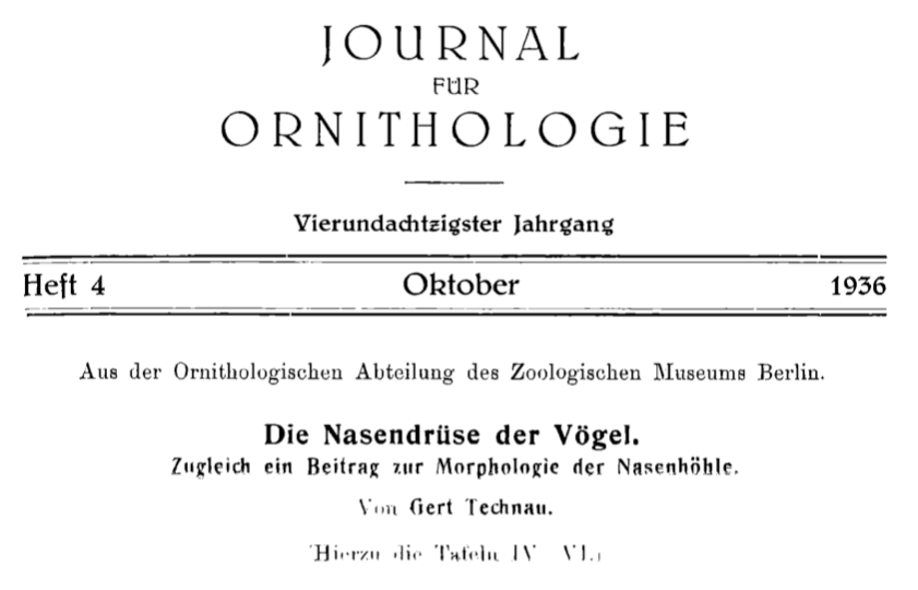

Gerhard Technau in Berlin made an extensive comparative study of the nasal glands of birds. His paper, published in 1936, is over 100 pages long. When, in 1975, we compared Technau’s findings with a list of birds known to have functional salt glands, we found that salt glands are not always in the supraorbital position. These early studies have been confirmed more recently. The salt gland in gannets, boobies, cormorants, shags, and pelicans, for example, are situated within the roof of the orbit; depressions in the bone are found there.

We used Technau's diagram for our book in 1975. We stuck with Technau's
nomenclature but the sharp-eyed will notice his terminology is incorrect.
His 'interorbital' (i.e. between the orbits) should be 'intraorbital' (within
the orbit).


Thus it is entirely possible that other early birds, as well as their dinosaur ancestors, had salt glands as well as those leaving evidence of supraorbital nasal glands in their fossilised remains.

The new paper is:

Wang X, Huang J, Hu Y,  Liu X, Peteya J, Clarke JA. 2018. The earliest evidence for a supraorbital salt gland in dinosaurs in new Early Cretaceous ornithurines. Scientific Reports 8:3969 | DOI:10.1038/s41598-018-22412-8. Note - two - and I have only checked two - of the references given in this paper are incorrect.

Other references

Technau G. 1936. Die Nasendrüse der Vögel. Zugleich ein Beitrag zur Morphologie der Nasenhöhle. Journal für Ornithologie 84, 511-617.
( I have been able to find nothing about Technau nor his other work.)

Siegel-Causey D. 1990. Phylogenetic patterns of size and shape of the nasal gland depression in Phalacrocoridae. The Auk 107, 110-118.

Marples BJ. Structure and development of nasal glands in birds. Proceedings of the Zoological Society of London (1932), 829-844.

Peaker M, Linzell JL. 1975. Salt Glands in Birds and Reptiles. Cambridge: Cambridge University Press.

*Relevant here is the recent work on using knowledge of the structure of nasal salt glands to infer what was happening in extinct birds has appeared so far only as an abstract:
Caggiano EG, Cerio D, Porter R, Ridgely RC, Witmer LM. 2017. The nasal salt gland of extant birds: anatomical structure and its relevance for inferring the behavior and habitat preferences of extinct birds. FASEB Journal 81, Abstract 579.5. 

Tuesday, 27 March 2018

Not a Giant Snake - Just a Broken Mollusc Shell. Sir John Graham Kerr’s Howler: How was the Misidentification Perpetuated?

Sir John Graham Kerr
A couple of weeks ago I was in the Graham Kerr Building at Glasgow University for a meeting. Still known to the old lags as ‘Zoology’, it is named for Sir John Graham Kerr FRS, Professor of Natural History and then, when the department was split, of Zoology from 1902 until 1935. The 1920s zoology building was very much his building and his empire.

A natural historian of the old school he is perhaps better known now for a horrendous misidentification of a specimen than remembered for his research, on lungfish, for example, for his wrong-headed—but not unusual for the time—views on evolutionary pathways, for his antipathy to experimental biology, for disruptive camouflage of ships, for his political life as a Member of Parliament for the Scottish Universities before such seats were abolished and for his pugnacity.

The misidentification arose from a find among a mass of bones obtained from the Gran Chaco of South America by his friend, the missionary Andrew Pride. Kerr identified the specimen as the fang of a snake and, given its size—nearly 6.5 cm measured on the outside of the curve—a giant snake to which he gave the name Bothrodon pridii which translates to Pride’s Furrow-toothed Snake. The paper was published in Proceedings of the Royal Society of Edinburgh in 1927.


From Kerr's 1927 paper

Kerr interpreted his fang as being from an opisthoglyphous snake like the Boomslang with the tooth serving to grasp the prey while the poison worked its way down the furrow and into the wound. He then calculated that the fang’s owner must have been about 60 feet long or more. A true piece of deduction from a single part of the anatomy that Richard Owen would have been proud of. Or would he?

Only after a cast of the ‘fang’ was sent to Dr Werner Quenstedt (1893-1960) in Berlin did it become apparent that far from being the tooth of a snake, Kerr’s specimen was the broken off projection from the shell of a Chiragra Spider Conch, now known as Harpago chiragra, from the Indo-Pacific. Oh dear.


Chiragra Spider Conch
from Wikipedia. Photograph by H. Zell

Quenstedt’s correction to Kerr’s identification was published in 1939. However, something odd in the what-happened-next category was noted in a blog post by Dr Karl Shuker. Graham Kerr continued to refer to the discovery of the tooth and to Bothrodon in his book published in 1950, A Naturalist in Gran Chaco. Shuker wonders whether Kerr simple did not accept that he was wrong.

The ‘Bothrodon pridii fang’ was clearly Kerr’s pride (if you will forgive the pun) and joy. Even after Quenstedt’s publication in 1939 Kerr demonstrated the specimen at a Royal Society Club dinner in 1943 (mistakenly reported as 1940 by others). But what is even odder is that Kerr’s successor (and relation by marriage) at Glasgow, Edward Hindle FRS (1886-1973) in a biographical memoir for the Royal Society written shortly after Kerr’s death in 1957 refers to the fact that the specimen was one of Kerr’s favourite exhibits in his departmental museum and praises Kerr for its identification. Hindle continued:
Unfortunately no other part of the skeleton has ever been found but comparing the size of this fang with that of a modern poisonous snake, it is estimated that this monster may well have been some 60 feet long, surely one of the most formidable animals that ever lived.

It is possible that Kerr did not accept Quenstedt’s debunking of his pet specimen as a misidentification. Or is there another explanation?


From Allibone (a book printed by early camera-ready technology)


The Holborn Restaurant at the corner of High Holborn and Kingsway.
Now demolished, this famous London restaurantwas used by the
Royal Society Club for dinners between 1942 and 1944.

Graham Kerr left Glasgow in 1935 for the political arena and scientific retirement. Is it just possible that nobody told him, nor dared tell him, what had happened in Berlin?

It is also possible that Quenstedt’s work was more widely known in the U.S.A. than in Britain. Even by 1939 standards the publication looks pretty obscure†. The outbreak of war 1939 saw contacts cut between German and British scientists. But until the U.S.A. eventually, as W.S. Churchill might have phrased it, declared war on Germany in 1941 after the Japanese attack on Pearl Harbour, some contact was maintained and publications exchanged.

Edward Hindle’s ignorance is harder to explain. During the 1940s and 50s Hindle was very much the man-about-town gentleman scientist in London. Even though no palaeontologist nor herpetologist it seems odd that he seemed unaware of what had happened as he flitted from this scientific society dinner to that learned society event while ensconced at London Zoo from 1943 until 1951 and the during his retirement. Even had he known of the misidentification and had wished to avoid Kerr, his late first wife’s cousin as well as his predecessor in Glasgow, being remembered for it he surely would have simply ignored the topic in the biographical memoir rather than listing it as an achievement.

Allibone TE. 1976. The Royal Society and its Dining Clubs. Oxford: Pergamon.

Hindle E. 1958. John Graham Kerr, 1869-1957. Biographical Memoirs of Fellows of the Royal Society 4, 155-166.

Kerr JG. 1927. Bothrodon pridii, an extinct serpent of gigantic dimensions. Proceedings of the Royal Society of Edinburgh 46, 314-315. A number of sources quote this paper as being published in 1926.

Kerr JG. 1950. A Naturalist in the Gran Chaco. Cambridge University Press.

†The reference is given as Quenstedt in Kuhn, Oskar. 1939. This must be: Kuhn O. 1939. Squamata : Lacertilia et Ophidia. Fossilium catalogus, 1 . Animalia / editus a W. Quenstedt ; pars 86. W. Junk.

Sunday, 25 March 2018

Porcupines in Hong Kong: Letters to the South China Morning Post in 1930

Further to my previous post on our night safari to see porcupines in Hong Kong, these are letters on the subject I found in the South China Morning Post of 21 November 1930.

Porcupines.
(To the Editor. S. C. M Post.) 
Sir,—In reply to the query by "A.B.". I hasten to assure him (or her) that porcupines are definitely among the native mammals of Hongkong island. Whether they are descendants of escaped animals brought from the mainland may be open to discussion but as long ago (or as recently) as 1927 it was established that they were breeding in the Shek-O neighbourhood, when one of them, in crossing the road, was run into by a motor-car.
     Late in 1928, while exploring the neighbourhood of the hillside above Tytam Tuk with a companion, we found definite traces of porcupines there.
V.

Sir,—In this morning's (Thursday's) issue of your paper "A.B.' mentions that he had seen, last week, a large porcupine on High West and asks whether these animals are natives of the Colony.
     Porcupines are native here and may occasionally be seen on the Peak, at Shekko, or in the New Territories.
     About 10 days ago, I purchased a porcupine which had been caught at Sha Tin; the animal had been raiding sweet-potato plantations and an all night watch had been set for it on three successive nights before its capture. This porcupine was a large healthy fellow but many of its quills had been shed, possibly during the excitement of its capture. Unfortunately, it escaped after only one night's sojourn in the University, and, after consuming various flowers and digging sundry holes, made off up the hillside.. Possibly, the fellow seen by “A.B.” is my porcupine.
     If anyone else sees "Fido," will they please treat the animal kindly, put some salt on his or her tail and lead him or her gently back to the University.
G. A. C. Herklots.
Herklots’s battles with porcupines raiding his garden in Pokfulam in 1947 were described in his The Hong Kong Countryside published in 1951, together with the problems caused by the quills removed from his garden raiders after their extermination:
The quills, distributed by my small daughter to her friends at school, proved popular at first but alas, the small boys finding from experience that they were very sharp used them with good effect on the small girls to their pain and discomfiture. Evidently porcupine quills should not be distributed in a co-educational school. D[epartment] of E[ducation] please note and instruct heads accordingly…

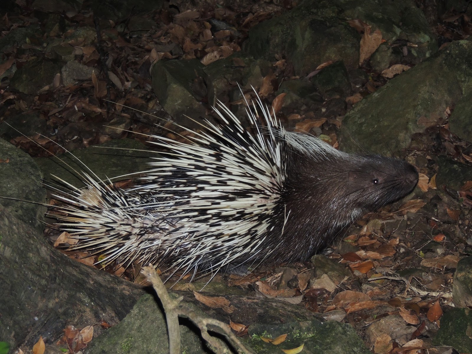
Malayan Porcupine in Hong Kong                                                                                                        [AJP Photo

Porcupines in Hong Kong: Our Night Safari

The Malayan or East-Asian Porcupine (Hystrix brachyura) is native to Hong Kong and has become much more common in recent decades than they were in the 1960s.

The paths of Hong Kong island are frequented by joggers in the evening. The joggers can get a surprise when a porcupine or sometimes a whole family appears in front of them.

Here is the result of our night safari at a location on Hong Kong island last November:


Friday, 23 March 2018

Monkey Hill at London: Monkey Temple at Bristol Zoo

There has been a great deal of recent interest in my article on Monkey Hill at London Zoo. In its postwar state it housed, for a time, Rhesus Macaques. Tuberculosis was blamed for the large number of deaths*. It was demolished in 1955.

Demolition of Monkey Hill
London Zoo
Children's Newspaper
2 April 1955

There were, though, other zoos in which groups of Rhesus Macaques were kept, apparently successfully. I can—just—remember going to Bristol Zoo at the age of three and seeing the Monkey Temple. I went there again around Easter 1963 and took this photograph. Bristol opened the Monkey Temple in 1928. It was apparently inspired by the Cold Lairs, an abandoned city inhabited by monkeys, in Rudyard Kipling’s The Jungle Book.

Monkey Temple, Bristol Zoo. My photograph from 1963

The years passed and such exhibits were—and still are—condemned by social and architectural historians with a political axe to grind as ‘colonial’, ’unnaturalistic’ or even ‘exotic’. 

I was highly amused when on my first visit to an Indian temple—in India—to see Rhesus Macaques disporting themselves on its walls and roof just as they did in Bristol. Far from being a colonial view of their natural habitat, that was their natural habitat!

The Monkey Temple in Bristol is now apparently used to house an exhibit on plants.

Postcard: Monkey Temple, Bristol (Clifton) Zoo


Monday, 19 March 2018

Arapaima ‘Milk’: Reflections on big fish and a big biological problem

Since the discovery of ‘lactation’ in discus fish, the production of mucus by a number of other species to feed their young has been described. Somewhat different has been the description of what happens in the Giant Arapaima (Arapaima gigas)—an endangered species of the Amazon basin and one that can reach a length of three metres. Locals had observed the gathering of young around the head of the male and the release from the head of a milky fluid into the water. Some work has been done on the phenomenon because these fish are bred and reared for human consumption.


Arapaima gigas captivity
Giant Arapaima (Credit: Citron / CC-BY-SA-3.0)

The fluid comes from the cephalic canals of the lateral line system. The eggs, larvae and young are in close contact with this secretion, even when the male is leading the offspring to water rich in plankton for feeding. Parental care last for three months. There are numerous possibilities for the function of “arapaima milk”. A paper published late last year* describes an attempt to set the ball rolling in this respect by taking a proteomic approach to the composition of the fluid.

The fluids from the cephalic canals breeding and non-breeding males and females were analysed since any additional substances present in the former could indicate a particular adaptation that benefits the young. To cut a long story short, the total protein content of the fluid was low, suggesting that the fluid is not important in nutrition, even if the young were to eat it. There were a number of antimicrobial agents in the vast array of peptides present. Two hormones, prolactin and stanniocalcin were detected in females.

The great problem, as with substances in mammalian milk, is to identify which, if any, substances of the hundreds present have a physiological rôle in the offspring. Fluid from the cephalic canals is bound to reflect cellular and protective systems within that system. They may be in the fluid for no other reason than they are disposable elements and have no function at all in the offspring. But protection for the cephalic canals against infection, which are open to the surrounding water, could simply being extended to provide some degree of protection to the offspring, from contact with the skin and gills and/or by ingestion.

The question of biological signals from parent to offspring through hormones or other biologically active substances has also been raised in discussions of skin or, in this case, cephalic secretions, of fish. For example, it might be tempting to speculate on whether the prolactin detected in female arapaima milk has any effect on the offspring. However, it is a hormone that one would expect to be present given its function in mediating aspects of maternal care in vertebrates and its presence in fluid could simply reflect an action on the tissues of the cephalic canals rather than any significance to the offspring.

Mammalian milk contains a large number of hormones, growth factors and the like which could have an effect on the young. So many people were—and still are—assuming that presence in milk indicates a function in the young mammal ingesting the milk that Peggy Neville and I warned against making such assumptions and published a list of criteria that would need to be met in order to demonstrate that such an effect was taking place:

  1. An effect in the offspring must be obtained in response to exposure to the substance in milk.
  2. The effect in the offspring must be abolished by removal of the substance from milk and restored when that specific component is restored.
  3. Th substance must be shown to be present and active in milk.
  4. The substance must be shown to retain its biological activity in the offspring to the point at which it is postulated to act or to be activated by partial digestion within the digestive tract.

Shortly after we wrote that commentary in 1991 only two series of studies satisfied those criteria.

The criteria for demonstrating an effect of a substance in milk on the offspring are tough to fulfil. But they have to be in order to prevent the proliferation of just-so stories masquerading as science. It would be a simple matter to modify those criteria to parental care in fish and any potential chemical communication between parent and offspring through skin or cephalic secretions.

The only clue I have seen so far for the importance of arapaima milk is: ‘Previous work suggested fingerlings raised under parental care condition would have higher survival rates and enhanced growth performance compared to in-door reared ones’*.

I also wondered—and have not seen discussed (although I have not looked that hard)—whether arapaima milk had some chemoattractant function, keeping the offspring concentrated around the head of the male when he finds plankton-rich but possibly murky water in which they can feed. But like every other suggestion that is a just-so story that ‘ain’t necessarily so’.

*Torati LS, Migaud H, Doherty MK, Siwy J, Mullen W, Mesquita PEC, Albalat A. 2017. Comparative proteome and peptidome analysis of the cephalic fluid secreted by Arapaima gigas (Teleostei: Osteoglossidae) during and outside parental care. PLoS ONE 12(10): e0186692. https://doi.org/ 10.1371/journal.pone.0186692

Peaker M, Neville MC. 1991. Hormones in milk: chemical signals to the offspring? Journal of Endocrinology 131, 1-3.

Saturday, 17 March 2018

Yellow-crested Cockatoo: critically endangered but still being traded illegally

In February 2017 I wrote a series of posts on the critically endangered Yellow-crested Cockatoo (Cacatua sulphurea) of Indonesia and East Timor. News this week from TRAFFIC shows that this cockatoo is still being caught and traded for the pet trade in south-east Asia. An illegal shipment of animals, including a hundred cockatoos of three species, was seized in the Philippines last week.